 Antibody Fluorescein Conjugated")

Datasheet is currently unavailable. Try again or CONTACT US
Mouse IgG (H&L) Antibody Fluorescein Conjugated
Goat Polyclonal
13 References
610-1202-0500
610-1202
500 µg
2 mg
Lyophilized
Lyophilized
WB, IHC, IF, Dot Blot, Multiplex
Mouse
Goat
Shipping info:
Find your nearest distributor for timely delivery and local support.
Product Details
Goat Anti-Mouse IgG (H&L) Antibody Fluorescein Conjugated - 610-1202
Goat Anti-Mouse IgG Secondary Antibody fluorescein Conjugated, Goat Anti-Mouse IgG Antibody FITC Conjugated, GAM-FITC, Anti-mouse IgG secondary antibody, anti-mouse IgG Fluorescein conjugated secondary antibody, Gt Anti-Ms IgG FITC Conjugated Antibody
Goat
IgG (H&L)
Fluorescein (FITC)
Polyclonal
IgG
2.75
Target Details
Mouse
Mouse IgG whole molecule
Goat Anti-Mouse IgG [H&L] FITC Conjugated Antibody was prepared from monospecific antiserum by immunoaffinity chromatography using Mouse IgG coupled to agarose beads followed by solid phase adsorption(s) to remove any unwanted reactivities. Assay by immunoelectrophoresis resulted in a single precipitin arc against anti-Fluorescein, anti-Goat Serum, Mouse IgG and Mouse Serum.
Application Details
Dot Blot, WB
IF, IHC, Multiplex
- View References
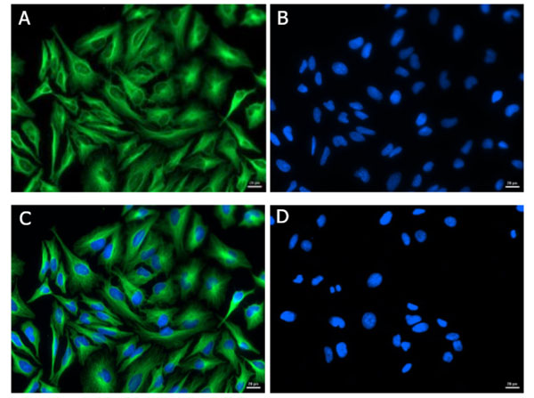
Anti-Mouse IgG FITC Conjugated Antibody has been tested by dot blot and western blot and is designed for immunofluorescence microscopy, flow cytometry, fluorescence based plate assays (FLISA) and fluorescent western blotting. This product is also suitable for multiplex analysis, including multicolor imaging, utilizing various commercial platforms.
Formulation
2.0 mg/mL by UV absorbance at 280 nm
0.02 M Potassium Phosphate, 0.15 M Sodium Chloride, pH 7.2
0.01% (w/v) Sodium Azide
0.1 mg/ml Bovine Serum Albumin (BSA) - IgG and Protease free, 50% (v/v) Glycerol
1.0 mL
Restore with deionized water (or equivalent)
Shipping & Handling
Ambient
Store vial at 4° C prior to restoration. For extended storage aliquot contents and freeze at -20° C or below. Avoid cycles of freezing and thawing. Centrifuge product if not completely clear after standing at room temperature. This product is stable for several weeks at 4° C as an undiluted liquid. Dilute only prior to immediate use.
Expiration date is one (1) year from date of receipt.
Anti-Mouse IgG [H&L] Fluorescein Antibody generated in goat detects reactivity to Mouse IgG. Secreted as part of the adaptive immune response by plasma B cells, immunoglobulin G constitutes 75% of serum immunoglobulins. Immunoglobulin G binds to viruses, bacteria, as well as fungi and facilitates their destruction or neutralization via agglutination (and thereby immobilizing them), activation of the compliment cascade, and opsinization for phagocytosis. The whole IgG molecule possesses both the F(c) region, recognized by high-affinity Fc receptor proteins, as well as the F(ab) region possessing the epitope-recognition site. Both the Heavy and Light chains of the antibody molecule are present. Secondary Antibodies are available in a variety of formats and conjugate types. When choosing a secondary antibody product, consideration must be given to species and immunoglobulin specificity, conjugate type, fragment and chain specificity, level of cross-reactivity, and host-species source and fragment composition.
Liu M et al. (2023). Co-culture models of endothelial cells, macrophages, and vascular smooth muscle cells for the study of the natural history of atherosclerosis. PLoS One.
Applications
WB, IB, PCA
Liu M et al. (2023). Role of triggering receptor expressed on myeloid cells-1 in the mechanotransduction signaling pathways that link low shear stress with inflammation. Sci Rep.
Applications
WB, IB, PCA
Kolijn D. et al. (2020). Enhanced Cardiomyocyte Function in Hypertensive Rats With Diastolic Dysfunction and Human Heart Failure Patients After Acute Treatment With Soluble Guanylyl Cyclase (sGC) Activator. Front Physiol.
Applications
IF, Confocal Microscopy; Multiplex Assay
Wang Y. et al. (2020). Exogenous spermine attenuates rat diabetic cardiomyopathy via suppressing ROS-p53 mediated downregulation of calcium-sensitive receptor. Redox Biol.
Applications
IF, Confocal Microscopy
Sundaramurthy S. et al. (2020). FHOD-1 is the only formin in Caenorhabditis elegans that promotes striated muscle growth and Z-line organization in a cell autonomous manner. Cytoskeleton (Hoboken).
Applications
IF, Confocal Microscopy
Saferali A, Tang AC, Strug LJ, et al. (2020). Immunomodulatory function of the cystic fibrosis modifier gene BPIFA1. PLoS One.
Applications
WB, IB, PCA
Vitacolonna M et al. (2017). In-vivo quantification of the revascularization of a human acellular dermis seeded with EPCs and MSCs in co-culture with fibroblasts and pericytes in the dorsal chamber model in pre-irradiated tissue. Cell Tissue Bank.
Applications
IF, Confocal Microscopy
Kryszke MH et al. (2016). Post-transcriptional gene silencing activity of human GIGYF2. Biochem Biophys Res Commun.
Applications
IF, Confocal Microscopy
Zhang K et al. (2015). Predicting Abdominal Aortic Aneurysm Target Genes by Level-2 Protein-Protein Interaction. PLoS One
Applications
IF, Confocal Microscopy
Adefolaju GA et al. (2015). BAX/BCL-2 mRNA and protein expression in human breast MCF-7 cells exposed to drug vehicles-methanol and dimethyl sulfoxide (DMSO) for 24 hrs. Niger Med J.
Applications
IHC, ICC, Histology
Cavaglia M et al. (2001). Regional variation in brain capillary density and vascular response to ischemia. Brain Res.
Applications
IHC, ICC, Histology
Bradley BJ et al. (1992). CD8 T cells are not required for islet destruction induced by a CD4+ islet-specific T-cell clone. Diabetes.
Applications
IF, Confocal Microscopy
SR Smith et al. (1990). Inhibition of collagen II-induced arthritis in mice--a comparison of the effects of Sch 24937, immunosuppressants and nonsteroidal antiinflammatory drugs on the clinical expression of disease. Int J Immunopharmacol.
Applications
IF, Confocal Microscopy
This product is for research use only and is not intended for therapeutic or diagnostic applications. Please contact a technical service representative for more information. All products of animal origin manufactured by Rockland Immunochemicals are derived from starting materials of North American origin. Collection was performed in United States Department of Agriculture (USDA) inspected facilities and all materials have been inspected and certified to be free of disease and suitable for exportation. All properties listed are typical characteristics and are not specifications. All suggestions and data are offered in good faith but without guarantee as conditions and methods of use of our products are beyond our control. All claims must be made within 30 days following the date of delivery. The prospective user must determine the suitability of our materials before adopting them on a commercial scale. Suggested uses of our products are not recommendations to use our products in violation of any patent or as a license under any patent of Rockland Immunochemicals, Inc. If you require a commercial license to use this material and do not have one, then return this material, unopened to: Rockland Inc., P.O. BOX 5199, Limerick, Pennsylvania, USA.